Vol. 1 (2020), Article ID 246105, 4 pages
Research Article
A Short-Period Therapy with Subcutaneous Low-Dose IL-2 Plus High-Dose Melatonin to Correct Advanced Cancer-Related Lymphocytopenia: Possible Impact on the Survival Time
Paolo Lissoni, Giusy Messina, and Giuseppe di Fede
Institute of Biological Medicine, 20129 Milan, Italy
Received 11 June 2020; Revised 3 September 2020; Accepted 2 October 2020; Published 19 October 2020
Paolo Lissoni, Giusy Messina, and Giuseppe di Fede, A Short-Period Therapy with Subcutaneous Low-Dose IL-2 Plus High-Dose Melatonin to Correct Advanced Cancer-Related Lymphocytopenia: Possible Impact on the Survival Time, Psychoneuroimmunology Journal, 1 (2020), art246105. doi:10.32371/pnij/246105
Despite the well-known importance of lymphocytes in tumor cell destruction and the evidence that lymphocytopenia is associated with a poor prognosis and lower survival in almost all tumor histotypes, lymphocyte count is not generally taken into consideration by the oncologists and no specific protocol has been proposed to treat cancer-related lymphocytopenia because of its negative impact on the clinical course of the neoplastic disease. Obviously, the most effective agents to stimulate the proliferation of lymphocytes would have to be their major growth factor, consisting of IL-2, whose in vivo biological activity may be further amplified by an association with some neuro-immunomodulating agents, such as the pineal hormone melatonin (MLT). This study was performed to evaluate the effects of subcutaneous (SC) low-dose IL-2 plus high-dose MLT in a group of metastatic cancer patients with persistent lymphocytopenia, who failed to respond to the standard anticancer therapies. The study included 14 patients who received MLT orally at 100 mg/day in the evening every day without interruption plus IL-2 SC at 1.8 MIU/day for five days a week for two consecutive weeks, corresponding to one complete cycle. A second cycle was repeated after a rest period of two weeks. A normalization of lymphocyte count was achieved in 9/14 (64%) patients, 4 of them (29%) just after the first week of therapy. Because of the association between cancer-related lymphocytopenia and lower survival and the fundament role of lymphocyte in mediating cancer cell destruction, the correction of advanced cancer-related lymphocytopenia by a short-term SC low-dose IL-2 therapy could improve the clinical course of cancer patients, including those suitable for the only palliative treatment.
cancer immunotherapy; interleukin-2; lymphocytopenia; melatonin
The failure of the antitumor immunity in advanced cancer patients does not depend only on a deficiency of T lymphocyte system, including T helper (Th) (CD4+) and cytotoxic T lymphocytes (CD8+) [1,2,3], but also on a concomitant enhanced activation of the macrophage system [4,5,6]. This is because the macrophage-induced chronic inflammation has appeared to allow a diminished Th and cytotoxic T lymphocyte functions by promoting regulatory T-cell (Treg) (CD4+CD25+) activation, which suppresses the antitumor immunity [3,4,5,6,7]. From a clinical point of view, recent clinical studies have shown that the simple lymphocyte-to-monocyte ratio (LMR) may synthetically reflect the status of lymphocyte-macrophage system interactions [8,9], since the evidence of an abnormally low LMR with values less than 2.1 has been proven to be associated with a poor prognosis and predicting a lower survival time in metastatic cancer patients. The inhibition of the macrophage system may be achieved by nonsteroidal anti-inflammatory agents, or more effectively by cannabinoid agents [10] and the pineal indole hormones; the most investigated of them is melatonin (MLT) [11,12,13]. In any case, it has been shown that the pineal is only the main source of MLT, but not the only one, since MLT may be produced by several other tissues, including the gastrointestinal tract, the gonads, and the lymphocytes themselves [11,12]. From a physiological point of view, the pineal constitutes the main connection between the universal conditions and the biological status of the single living organisms by acting
as a neurochemical transducer and influencing the overall biological functions, including DNA expression, in relation to the different environmental conditions [12]. On the other side, the most active endogenous agent to stimulate T lymphocyte proliferation is their own growth factor, IL-2, which in fact was defined as a T-cell growth factor (TCGF) [1]. Moreover, in addition to its inhibitory effects on macrophage-mediated inflammatory response, MLT has also been proven to directly stimulate Th cell proliferation and IL-2 secretion by acting on specific MLT receptors expressed by lymphocytes themselves. Then, lymphocyte count could be enhanced through an immune way by the administration of cytokines such as IL-2 and, namely, IL-2 itself, or alternatively through a neuroimmune approach with molecules provided by neuro-immunomodulating properties of which the most investigated is the pineal hormone MLT, which may be defined as a neuroendocrine immunomodulating agent. In fact, previous preliminary studies had already demonstrated the greater efficacy of IL-2 plus MLT, compared with IL-2 alone, in enhancing the number of lymphocytes in cancer patients [14]. This finding is not surprising, since MLT could enhance IL-2 efficacy either by amplifying its stimulatory effect on lymphocyte proliferation or reducing the potentially negative stimulatory action of IL-2 also on the macrophage and Treg systems which in contrast inhibit the antitumor immunity. Then, the neuro-immunotherapeutic schedule of subcutaneous (SC) low-dose IL-2 and high-dose MLT could constitute the optimal nontoxic regimen to enhance lymphocyte count in advanced cancer patients. The present preliminary phase-II clinical study was performed in an attempt to start to define the optimal neuroimmune schedule of IL-2 and MLT administration to correct cancer-related lymphocytopenia as well as to establish the time required to normalize lymphocyte count and the duration of lymphocyte normalization in metastatic cancer patients, for whom no other standard anticancer therapy was available, and then suitable only for the best supportive care.
The phase-II study included 14 consecutive untreatable metastatic cancer patients (M/F: 6/8; median age: 61 years, range 49–71), who underwent only palliative treatments because of lack of response to previous standard antitumor therapies. After the approval of the Ethical Committee, the experimental protocol was explained to each patient and a written consent was obtained. The immunotherapeutic cycle consisted of SC IL-2 at 1.8 MIU/day in the afternoon for five days a week for two consecutive weeks in association with MLT at 100 mg/day orally during the dark period of the day; every day without interruption; starting seven days prior to IL-2. A second cycle was planned after a two-week rest period, and a further third cycle was programmed only in patients who did not show any clear increase in lymphocyte count in response to the previous two cycles. Eligibility criteria were as follows: histologically proven metastatic solid tumor, measurable lesions, persistent lymphocytopenia with lymphocyte count less than 1,000/mm3 for at least three months prior to the study, no availability of possible effective standard anticancer therapies because of lack of response to the commonly used anticancer therapies (including chemotherapy, endocrine therapy, and targeted therapies) or very poor clinical conditions, which make the patients as unable to tolerate the standard anticancer treatments, no concomitant autoimmune disease, and no chronic therapy with corticosteroids because of their potential lympho-cytotoxic activity. Patients with brain metastases were excluded from the study, because of the possible IL-2-induced increase in cerebral oedema. Tumor histotypes were as follows: lung adenocarcinoma: 3; colorectal cancer: 3; pancreatic adenocarcinoma: 2; gynaecologic tumors: 2; malignant melanoma: 1; renal cell cancer: 1; hepato-carcinoma: 1; soft tissue sarcoma: 1. Lymphocyte count and LMR values were determined prior to therapy and at 5-day intervals for the whole period of IL-2 injection, and at 15-day intervals for the three months after the interruption of treatment. Data were statistically evaluated by the chi-square test (ANOVA) and the Student’s t-test, as appropriate to evaluate the timing of improvements in immune cell numbers.
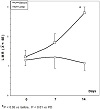
A normalization of lymphocyte number with values greater than 1,500/mm3 was achieved in 9/14 (64%) patients, among them 4/14 (29%) patients obtained lymphocyte normalization just after the first week of the first cycleof treatment, 3 other patients (21%) after the first cycle of therapy, and the last 2 patients (14%) after two cycles of therapy, whereas no further increase was observed after three cycles of therapy in patients who did not achieve any lymphocyte rise after the first two cycles of treatment. Changes in mean values of lymphocytes and monocytes occurring on therapy were illustrated in Figure 1. In responder patients, lymphocyte and monocyte mean numbers significantly increased (P < .01 vs. before) and decreased (P < .05 vs. before) on treatment, respectively. On the contrary, in non-responder patients no significant changes in lymphocyte mean number occurred, whereas monocyte mean count increased on treatment, even though without significant differences with respect to the pretreatment values. Before therapy, lymphocyte count was below 1,000/mm3 in 6 patients, whereas it was ranging between 1,000 and 1,500/mm3 in the remaining 8 patients. Moreover, as illustrated in Figure 2, lymphocyte increase was significantly greater in patients with lymphocyte count ranging from 1,000 to 1,500 prior to therapy than in those with values less than 1,000/mm3 (P < .05), whereas no difference was seen in the behavior of monocytes. After three months of therapy, lymphocyte count still persisted to be higher than 1,500/mm3 in 6/9 (675) patients who achieved a normalization of lymphocyte count on treatment. Finally, all patients showed abnormally low pretreatment LMR values. A normalization of LMR with values more than 2.1 on therapy was achieved in 8/14 (57%) patients, without significant differences between patients with lymphocyte count less than 1,500/mm3 and those with values less than 1,000/mm3 prior to therapy (5/8 (63%) vs. 3/6(50%)). Data were normally distributed and of equal variance. No important toxicity was observed on treatment, and in particular no relevant hypotension occurred on study. Fever less than 38 °C was observed in 5/14 (36%) patients. Asthenia was referred by 7/14 (50%) patients, while myalgia occurred only in 4/14 (29%) patients.

Figure 1: Lymphocyte, monocyte, and lymphocyte-to-monocyte ratio (LMR) under IL2.

Figure 2: LMR under IL2 in patients with stable disease (SD) or progressive disease (PD).
The present preliminary phase-II study shows that a neuroimmunotherapeutic schedule with a short period of SC low-dose IL-2 plus high-dose MLT is sufficient to correct advanced-cancer related lymphocytopenia in most patients. Moreover, the study seems to show a greater efficacy of treatment in patients with no important monocyte increase on treatment. This evidence would suggest that the effect of IL-2 on lymphocyte number may depend also on its concomitant effect on monocyte count, which has been proven to reflect the activity of the macrophage system and macrophage-tumor infiltration, which has appeared to be associated with a poor prognosis in cancer patients [4,5,6,7]. Until now, IL-2 was used as an anticancer agent to induce a destruction of cancer cells. However, because of its ability to stimulate lymphocyte proliferation, this study would suggest that IL-2 may be also used to indirectly influencing tumor progression by correcting cancer-related lymphocytopenia, since the evidence of low lymphocyte count has been proven to predict a poor prognosis in advanced cancer patients [15]. Obviously, because of the different individual biological response to therapy, different IL-2 dosages and timing of injection will be required for each patient by monitoring across the time its lymphocyte behavior. The lack of lymphocyte response to IL-2 observed in some patients could be due to the presence of several other cytokine alterations, including IL-12 deficiency and high levels of inflammatory cytokines (such as IL-6, IL-1 beta) or immunosuppressive cytokines (such as IL-10 and TGF-beta). Therefore, the measurement of the most important cytokines involved in the control of tumor progression will be required to explain the lack of lymphocyte response to IL-2 in advanced cancer patients. Moreover, further studies will be needed to evaluate the impact of lymphocytopenia correction on the survival time of metastatic cancer patients, for whom no other standard antitumor therapies are available, in an attempt to propose a potentially effective therapy also to patients considered as suitable for the only palliative therapy. Finally, further clinical researches with more adequate measures will be needed to better establish the influence of IL-2 plus MLT immunotherapy on the quality of life of cancer patients. In any case, it is important to remember that IL-2 immunotherapy, which historically was the first cancer immunotherapy founded not only on empiric evidences, but on specific immune mechanisms [1], was clinically progressively abandoned after the demonstration of its potential stimulatory action on Treg lymphocytes [16], which in contrast suppress the anticancer immunity [3,7]. However, successive studies have shown that in addition to the protumoral action of Treg-related TGF-beta, another cytokine, IL-17, may play an important stimulatory action on cancer growth [17], as confirmed by the evidence that IL-17 expression is associated with a greater malignancy of most tumor histotypes [18,19]. IL-17 inhibits TGF-beta secretion [20], which promotes cancer growth, but the protumoral action of IL-17 has been proven to be due to a direct stimulatory effect on cancer cells proliferation [17,18,19] and a stimulation of the macrophage system [21]. IL-2 has appeared to be able to inhibit IL-17 secretion from Th17 lymphocytes [21,22]. Then, because of the inhibitory action of IL-17 on TGF-beta secretion [20], IL-2-induced stimulation of Treg cells and TGF-beta secretion could simply represent the consequence of the inhibitory effect of IL-2 on IL-17 secretion. Therefore, in addition to its role as T lymphocyte growth factor, IL-2 could be successfully reintroduced in the clinical oncology, despite its potential stimulatory action of Treg cells, in an attempt to counteract IL-17 secretion, whose role in cancer progression seems to be fundamental, by representing one of the main endogenous protumoral molecules [17,18,19].
The authors declare that they have no conflict of interest.
- E. A. Grimm, A. Mazumder, H. Z. Zhang, and S. A. Rosenberg, Lymphokine-activated killer cell phenomenon. Lysis of natural killer-resistant fresh solid tumor cells by interleukin 2-activated autologous human peripheral blood lymphocytes, J Exp Med, 155 (1982), 1823–1841.
- P. Lissoni, Prognostic markers in interleukin-2 therapy, Cancer
Biother Radiopharm, 11 (1996), 285–287.
- W. Zou, Regulatory T cells, tumour immunity and immunotherapy, Nat Rev Immunol, 6 (2006), 295–307.
- A. Mantovani, P. Allavena, A. Sica, and F. Balkwill, Cancer-related
inflammation, Nature, 454 (2008), 436–444.
- S. I. Grivennikov, F. R. Greten, and M. Karin, Immunity,
inflammation, and cancer, Cell, 140 (2010), 883–899.
- P. Lissoni, Therapy implications of the role of interleukin-2 in cancer, Expert Rev Clin Immunol, 13 (2017), 491–498.
- R. Kim, M. Emi, K. Tanabe, Y. Uchida, and T. Toge, The role of Fas ligand and transforming growth factor beta in tumor progression: molecular mechanisms of immune privilege via Fas-mediated apoptosis and potential targets for cancer therapy, Cancer, 100 (2004), 2281–2291.
- L. Gu, H. Li, L. Chen, X. Ma, X. Li, Y. Gao, et al., Prognostic role of lymphocyte to monocyte ratio for patients with cancer: evidence from a systematic review and meta-analysis, Oncotarget, 7 (2016), 31926–31942.
- P. Lissoni, G. Messina, F. Rovelli, L. Vigorè, A. Lissoni, and G. Di Fede, Low lymphocyte-to-monocyte ratio is associated with an enhanced regulatory T lymphocyte function in metastatic cancer patients, Int J Recent Adv Multidiscip Res, 5 (2018), 3353–3356.
- F. Grotenhermen, Pharmacology of cannabinoids, Neuro Endocrinol Lett, 25 (2004), 14–23.
- G. J. Maestroni, The immunoneuroendocrine role of melatonin, J Pineal Res, 14 (1993), 1–10.
- A. Brzezinski, Melatonin in humans, N Engl J Med, 336 (1997), 186–195.
- P. Lissoni, The pineal gland as a central regulator of cytokine network, Neuro Endocrinol Lett, 20 (1999), 343–349.
- P. Lissoni, S. Barni, G. Tancini, A. Ardizzoia, G. Ricci, and R. Aldeghi, A randomised study with subcutaneous low-dose interleukin 2 alone vs interleukin 2 plus the pineal neurohormone melatonin in advanced solid neoplasms other than renal cancer and melanoma, Br J Cancer, 69 (1994), 196–199.
- A. Riesco, Five-year cancer cure: relation to total amount of peripheral lymphocytes and neutrophils, Cancer, 25 (1970), 135–140.
- T. R. Malek, The main function of IL-2 is to promote the development
of T regulatory cells, J Leukoc Biol, 74 (2003), 961–965.
- L. Wang, T. Yi, M. Kortylewski, D. M. Pardoll, D. Zeng, and H. Yu, IL-17 can promote tumor growth through an IL-6-Stat3 signaling pathway, J Exp Med, 206 (2009), 1457–1464.
- S. Wang, Z. Li, and G. Hu, Prognostic role of intratumoral IL-17A expression by immunohistochemistry in solid tumors: a meta-analysis, Oncotarget, 8 (2017), 66382–66391.
- S. H. Chang, T helper 17 (Th17) cells and interleukin-17 (IL-17) in cancer, Arch Pharm Res, 42 (2019), 549–559.
- J. Lohr, B. Knoechel, J. J. Wang, A. V. Villarino, and A. K. Abbas, Role of IL-17 and regulatory T lymphocytes in a systemic autoimmune disease, J Exp Med, 203 (2006), 2785–2791.
- I. Kryczek, S. Wei, L. Vatan, J. Escara-Wilke, W. Szeliga, E. T. Keller, et al., Cutting edge: opposite effects of IL-1 and IL-2 on the regulation of IL-17+ T cell pool IL-1 subverts IL-2-mediated suppression, J Immunol, 179 (2007), 1423–1426.
- A. Laurence, C. M. Tato, T. S. Davidson, Y. Kanno, Z. Chen, Z. Yao, et al., Interleukin-2 signaling via STAT5 constrains T helper 17 cell generation, Immunity, 26 (2007), 371–381.
Copyright © 2020 Paolo Lissoni et al. This is an open access article distributed under the terms of the Creative Commons Attribution License, which permits unrestricted use, distribution, and reproduction in any medium, provided the original work is properly cited.